Prostate cancer is the second most commonly diagnosed cancer in men. Conventional screening relies on prostate specific antigen (PSA) levels in the blood, which can indicate prostate abnormalities but lacks specificity. Clinical examination and ultrasound also have limited sensitivity and often fail to detect early-stage tumours or to distinguish aggressive from indolent disease. Recently, multiparametric magnetic resonance imaging (mpMRI) has emerged as a more precise tool for detecting and assessing prostate cancer. It combines multiple imaging techniques to provide detailed information on tumour location, size and likelihood of malignancy. However, interpreting these complex scans requires specialised training, is subject to variability between clinicians, and remains a time-consuming process.
Harnessing the power of AI
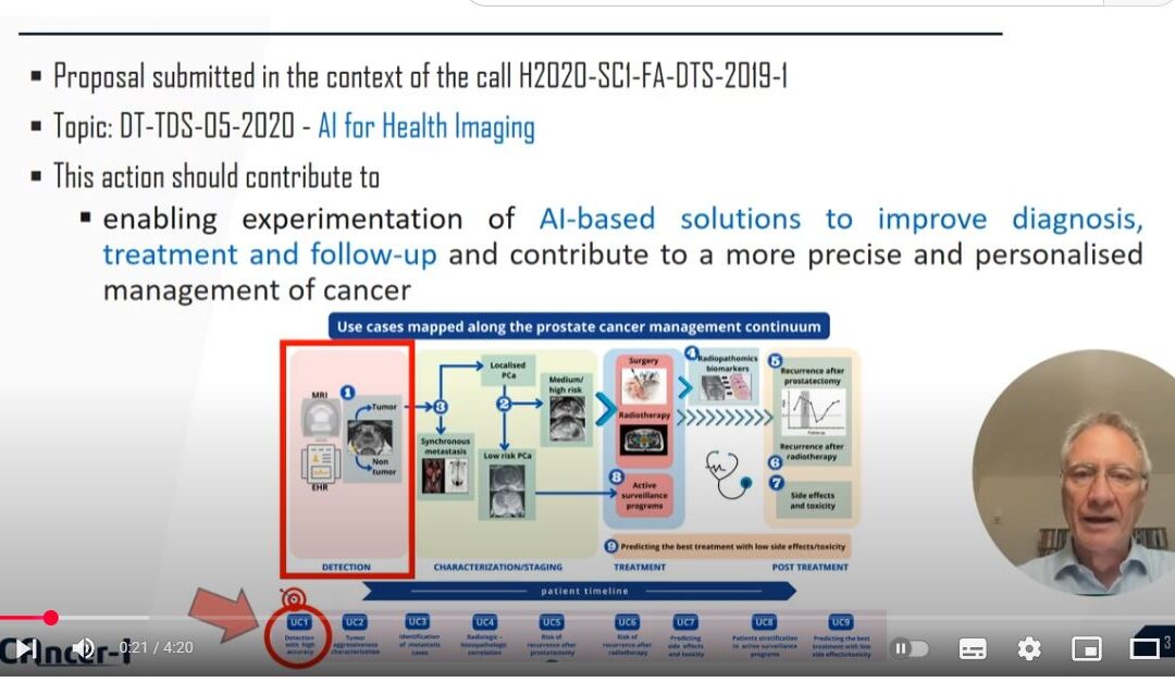
To address these challenges, the EU-funded ProCAncer-I(opens in new window) project has developed an AI-based platform that combines high-quality imaging data with clinical information to improve diagnosis, patient stratification and disease monitoring. “Our ambition was to develop AI models that could detect and characterise prostate cancer based on mpMRI scans, with higher performance than current standards,” states project coordinator Manolis Tsiknakis from the Foundation for Research and Technology-Hellas(opens in new window).
New standards in AI diagnostics
The ProCAncer-I platform is built on a large collection of prostate cancer imaging data. Retrospective and prospective data from over 14 000 patients and more than 9 million MRI images were used as a resource for developing AI algorithms tailored to real-world clinical needs. AI tools developed through the project include models for detecting cancer in both peripheral and transitional zones of the prostate, as well as characterising tumour aggressiveness and supporting lesion segmentation. These tools were tested in multicentre studies and showed clinical-grade accuracy.
Trustworthy AI
From the start, the project placed a strong emphasis on building trustworthy AI. With the project being one of the founders of the FUTURE-AI(opens in new window) initiative, significant effort was devoted to the design and development of tools and approaches that can ensure compliance with the FUTURE-AI guidelines. “We developed a novel AI model passport system that documents how each model was trained, validated and tested, making the entire process transparent and reproducible,” explains Tsiknakis. This includes tracking of data acquisition and pre-processing, model training and evaluation. In addition, the project created monitoring tools to detect when models might begin to underperform, allowing for timely intervention and quality assurance in clinical environments.
Clinical implementation
The ProCAncer-I models consider a wide array of imaging and non-imaging variables, such as PSA levels, lesion location and PIRADS scoring(opens in new window). This enables clinicians to make more objective risk assessment, detect aggressive cancers earlier, as well as select the best treatment for each patient. The several datasets that were generated, each responding to a specific clinical use case, have been integrated through a federated node into the European Cancer Imaging Initiative(opens in new window) contributing to the pan-European cancer imaging ecosystem. This ensures sustainability and alignment with broader European initiatives on health. Beyond its technical achievements, ProCAncer-I is expected to contribute to policy development. Looking ahead, the team aims to refine the predictive models of the platform and deepen collaboration with healthcare providers across Europe. “ProCAncer-I has shown that AI can be both powerful and responsible, helping clinicians with diagnosis,” concludes Tsiknakis.
Source


